PEMF Origin Story - Use for Treating Bone Fractures
Keywords - PEMF, Osteoporosis, Piezoelectricity/Voltage/Electricity, Currents of Injury, Reverse Piezoelectric Effect/Phonons/Resonators/Mechanical or Acoustic Waves, Crystalline Nature of Body, Wolff's Law, Regeneration/Stem Cells.
Video Series
History PEMF and Bones Fractures
Osteoporosis
iMRS 2000 Protocols
(themes - PEMF works, more is not better, low frequency and rapid rise and fall).
Should you fracture a bone in an arm or leg, and it fails to heal in 3-6 months, there is a good chance that your orthopedic surgeon will prescribe a PEMF device that will be placed near the fracture.
PEMF therapy is a safe and effective way of enhancing BONE formation. In fact this was the first medical and regulatory accepted uses for PEMF particularly in the treatment of non-union bone fractures. But not just non-union fractures and bone breaks, the SAME benefits and mechanisms apply to Osteopenia and Osteoporosis... In fact personally I think a low frequency and low intensity PEMF device is ESSENTIAL for anyone having bone loss issues or fractures of any kind (whether fresh, delayed, stress, or nonunion).
PEMF is the Ultimate bone building tool ALONG with strength training/vibration therapy and proper bone building supplements.
What the research for PEMF for bones SHOWS is that we do NOT need high intensity, the KEY is frequency resonance and a rapid rise and fall signal like a sawtooth! This sawtooth was and still is the MAIN waveform used for FDA approved PEMF devices for Bone fractures!
Lets first follow a brief review of the history of PEMF therapy that began a strong resurgence of electromedicine in the 1960s and 1970s!

Electrotherapy and energy medicine was actually thriving in the 1800's and very early 1900's. Both electricity and magnetism were used to heal bones, tissues and diseases of all kinds.
For example in the 1800s, physicians in London discovered that passing an electric current through the fracture site in a broken bone that did not heal could jump start the healing process. This became the method of choice for delayed union of fracture.
Magnetic therapy as a branch of medicine and as an area of clinical research fell into disfavor in North America as a result of the Abraham Flexner’s report on “Medical Education in the United States and Canada,” released in 1910.
The mail-order catalogues were purged of electromagnetic and other devices. Pharmaceutical companies proliferated and profited. Antibiotics, vaccines, drugs, surgery, xrays, etc.
The rise in power of political medicine and the shift to nearly 100% dependence on pharmaceuticals for health led to an unfortunate period of dormancy in the U.S., lasting 60 years, until the mid 1970s. It was then discovered that weak magnetic fields could induce sufficient current flow through the fracture to start the healing. The use of PEMF for bone healing was really the modern beginning of the Rise of PEMF therapy as one of the most power tools for healing not just bones, but everything from A-Z.
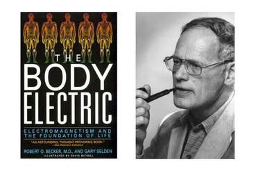
Though there were many figures in this resurgence, perhaps none was greater than the work of Robert O Becker that Led to PEMF
For example in the 1800s, physicians in London discovered that passing an electric current through the fracture site in a broken bone that did not heal could jump start the healing process. This became the method of choice for delayed union of fracture.
Magnetic therapy as a branch of medicine and as an area of clinical research fell into disfavor in North America as a result of the Abraham Flexner’s report on “Medical Education in the United States and Canada,” released in 1910.
The mail-order catalogues were purged of electromagnetic and other devices. Pharmaceutical companies proliferated and profited. Antibiotics, vaccines, drugs, surgery, xrays, etc.
The rise in power of political medicine and the shift to nearly 100% dependence on pharmaceuticals for health led to an unfortunate period of dormancy in the U.S., lasting 60 years, until the mid 1970s. It was then discovered that weak magnetic fields could induce sufficient current flow through the fracture to start the healing. The use of PEMF for bone healing was really the modern beginning of the Rise of PEMF therapy as one of the most power tools for healing not just bones, but everything from A-Z.
Though there were many figures in this resurgence, perhaps none was greater than the work of Robert O Becker that Led to PEMF
PEMF - The Ultimate Microcurrent Device
Robert Becker became interested in the fact that if a human loses a piece of bone, you will make more bone. But if you lose a piece of any other tissue it is replaced by scar tissue.
In humans it was once believed that only bone is capable of regeneration. We now know that all tissue can regenerate with the proper conditions.
Regeneration is the ability of lower animals to replace missing body parts. It is particularly evident in a salamander. The salamander is capable of growing an exact replacement of an arm, leg, eye, ear, up to 1/3 its brain, almost all its digestive tract and up to half its heart. If you poke a salamanders eye out, it will grow a new one. If you cut off his arm, he simply grows another one. The question is, "Why can the salamander grow new parts when frogs and we can't?
(The salamander has essentially the same anatomy as humans in that it has the same number of bones, muscles and nerves in the same arrangement.) The salamander is so effecient at regeneration that it does not get cancer.
The salamanders have a MUCH LARGER electrical potential OR Voltage. This voltage is energy that can be used to create a new limb... That was the only apparent difference between the frogs and salamanders, was that salamanders had a much stronger VOLTAGE which results in healing microcurrents and energy production (ATP). VOLTAGE is energy.
Becker surgically amputated the arms of salamanders and frogs and then used electrodes to measure the electrical potential at the point of tissue healing.
Becker wondered if delivering extra voltage to the frogs would do anything. When he did, and to his surprise, the FROGS GREW an entirely new limb!
Becker and his colleagues linked the capacity for regeneration to a direct current (DC) electric field, which can be found in all organisms. When a part of the body is cut or amputated, electrical currents are found to issue from the wound (as though from the ends of live wires in a circuit that have been cut).
These electrical currents are crucial for the processes of healing and regeneration.
One reason most organisms, like human beings, do not normally regenerate amputated parts is because the wound heals over too quickly, so the electrical currents stop flowing before the regenerate influences are complete. Successful regeneration was indeed reported for a child's lost fingertip by keeping the wound open until the regeneration was well on its way.
Jerry Tenant MD wrote a book called “Healing is Voltage” In his book he explains that injured / damaged cells require 2x more energy to repair.
----
The stimulus which initiates the complex regenerative process in amphibians has been reported to be a specific type of electrical signal, but the mechanism which provides the blueprint for the tissues to be regenerated is largely unknown.
Healing in general is known to be related to the degree of the injury, the amount of nerve tissue present at the site, and the electrical potential difference between the site and surrounding intact tissue (the "current of injury"). In particular, regeneration in amphibians such as salamanders and fracture healing in mammals are associated with complex changes in the local DC (direct current) electric field. An injury results in changes in the electric field and stimulates the animal's neural system, which in turn produces an electrical signal at the site of the injury, stimulating the complex cellular responses that eventually produce healing. The electric field gradually returns to normal, pre-injury levels as the injury heals. Conversely, failure of the normal healing process, as in fracture nonunions, is associated with the absence of appropriate electrical signals at the site of the injury.
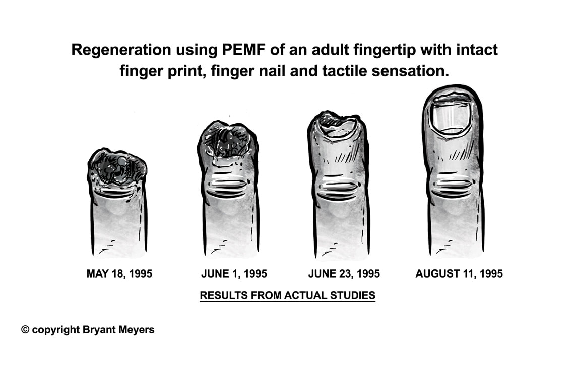
Robert O. Becker: Complete in-vitro regeneration of adult fingertip with intact finger print, finger nail and tactile sensation.
These electrical currents are crucial for the processes of healing and regeneration.
One reason most organisms, like human beings, do not normally regenerate amputated parts is because the wound heals over too quickly, so the electrical currents stop flowing before the regenerate influences are complete. Successful regeneration was indeed reported for a child's lost fingertip by keeping the wound open until the regeneration was well on its way.
Jerry Tenant MD wrote a book called “Healing is Voltage” In his book he explains that injured / damaged cells require 2x more energy to repair.
----
The stimulus which initiates the complex regenerative process in amphibians has been reported to be a specific type of electrical signal, but the mechanism which provides the blueprint for the tissues to be regenerated is largely unknown.
Healing in general is known to be related to the degree of the injury, the amount of nerve tissue present at the site, and the electrical potential difference between the site and surrounding intact tissue (the "current of injury"). In particular, regeneration in amphibians such as salamanders and fracture healing in mammals are associated with complex changes in the local DC (direct current) electric field. An injury results in changes in the electric field and stimulates the animal's neural system, which in turn produces an electrical signal at the site of the injury, stimulating the complex cellular responses that eventually produce healing. The electric field gradually returns to normal, pre-injury levels as the injury heals. Conversely, failure of the normal healing process, as in fracture nonunions, is associated with the absence of appropriate electrical signals at the site of the injury.
Robert O. Becker: Complete in-vitro regeneration of adult fingertip with intact finger print, finger nail and tactile sensation.
|
***Amputated Salamander: stump would become electropositive +25 mV (electron stealer) and he called this the "current of injury". The skin would grow over the stump and the cut ends in the nerve would connect with each skin cell called Neuroepithelial junction (NEJ). As soon as the NEJ forms, the reversed polarity changes normal cells to adult stem cells. So as soon as stem cells are formed Voltage goes to - 30 mV. During this time stem cells PROGRAMMED to what they are to become. Then Blastema forms and then current comes TO The injured area - current of healing - and arm rebuilds).
Most scientists do not believe that cells can differentiate from normal cells back to adult stem cells. It is believed that once cells differentiate into functional cells, they cannot go back the other way. |
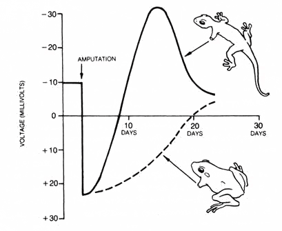
Electric potential changes at the cut end of the stump after amputation in salamander (top), which regenerates its amputated limb, and frog, which does not.
|
Regeneration vs Prosthetics for Bones (Example Hip)
Prosthesis - to add or put onto
a device, either external or implanted, that substitutes for or supplements a missing or defective part of the body.
1) Hip Replacements
2) Knee Replacement
3) Shoulder Replacement
4) Spinal Fusions and Back Surgery
5) Fractures that do not heal
Robert O Becker pointed out repeatedly that regeneration of damaged or missing body parts a far more natural and effective medical technology than the use of prosthetic devices or transplants.
Prosthetics fail to take into consideration
1) The human skeleton system is capable of considerable self-repair
2) No inorganic implant has the capacity of growth and remodeling and can ONLY decline in mechanical strength with time.
The statement that the best replacement for a damaged femoral head would be a new structure regenerated by the individual himself!
Becker has shown that very low levels of electrical stimulation can cause cultures of so called differentiated cells to de-differentiate into totipotent stem cells capable of form all of the tissues needed to replace a lost or damaged part.
The KEY to Becker's demonstration was reducing the strength of electrical stimulation. Following the usual way scientists tend to look at such matters, he assumed a large current would be more effective than a small one. The OPPOSITE was correct. In the experiments on frog red blood cells, conducted with a student named Frederick Brown, the test current was reduced a step at a time, until they reached the lowest level - a billionth of an ampere. THIS incredibly low level current produced a dramatic de-differentiation.
MORE IS NOT BETTER - The Extremely low levels of stimulation required to produce dedifferentiation and regeneration are comparible to the levels in the body and fields of the earth!
Surgeries and artificial parts are painful, expensive and riskly... the BETTER solution is more natural approaches of regeneration LIKE PEMF!!!
***Now, James Godwin and his colleagues have shown that macrophages are essential for salamanders' superherolike ability to sprout new limbs. The researchers studied the biochemical processes that occurred in salamanders at the site of a limb amputation. They then wiped out some or all of the macrophage cells to determine whether these cells were essential for regrowing the limbs.
Signals of inflammation were detected at the wound sites within one day of the amputations. Unexpectedly, anti-inflammatory signals, which normally arrive later in mammals recovering from injury, were also present at that time. Along with these signals, the researchers detected macrophages at the wound, peaking in number around four to six days after the injury.
The findings were detailed (May 20, 2013) in the journal Proceedings of the National Academy of Sciences.
In a total hip replacement (also called total hip arthroplasty), the damaged bone and cartilage is removed and replaced with prosthetic components. The damaged femoral head is removed and replaced with a metal stem that is placed into the hollow center of the femur.
|
|
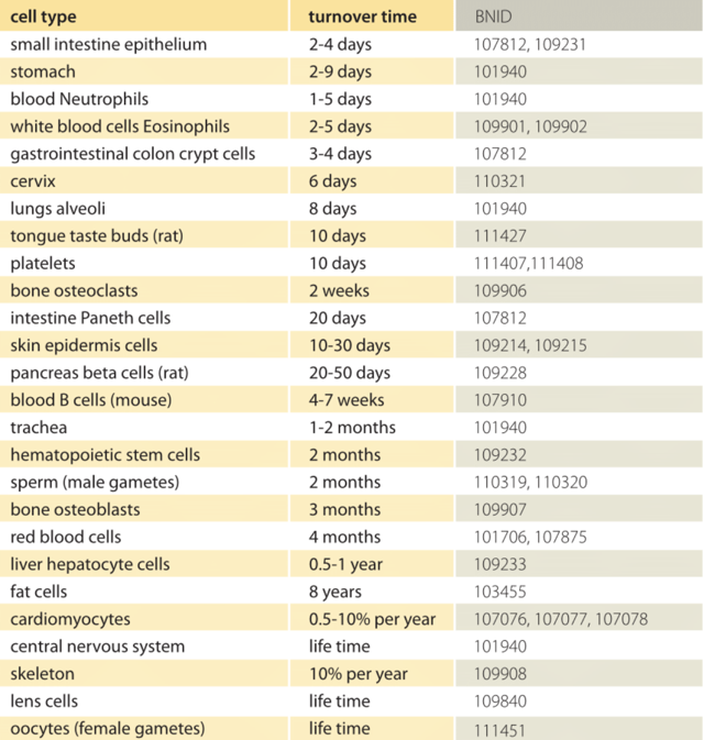
Normal Cell Regeneration Times in Humans
Source
http://book.bionumbers.org/how-quickly-do-different-cells-in-the-body-replace-themselves/
Source
http://book.bionumbers.org/how-quickly-do-different-cells-in-the-body-replace-themselves/
In 1961, Robert O Becker and Orthopedic Surgeon in New York gave a talk at an orthopedic meeting revealing on this very topic of regeneration
Andrew Basset was at this talk and met Becker and did work together investigating the electrical properities of Bone (piezo electric).
The significant research Findings of Dr Becker and Andrew Bassett have resulted in the widespread use of PEMF for Healing Fractured Bones.
Originally bone electrodes were used and then Bassett developed PEMF that induces the healing currents WITHOUT the need of invasive electrodes.
Bassett --> PEMF induces healing current flows at the molecular, cellular and tissue levels that are incredibly therapeutic.
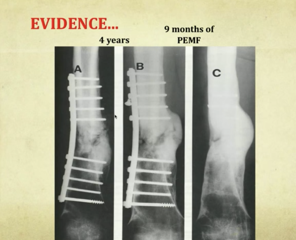
Bassett and others demonstrated that fracture non-unions could be stimulated to heal using tiny electric and magnetic fields. In his last scientific paper, orthopedic surgeon and medical researcher C.A.L. Bassett explained:
Jump starting a car with a dead battery creates an operation machine; exposure of a nonunion to PEMF can convert a stalled healing process to active repair, even in patients unhealed for as long as 40 years! (Bassett 1995)
The scientific evidence is that PEMF therapy is effective because it conveys 'information' that triggers specific repair activities within the body. The currents induced in tissues by PEMF mimic the natural electrical activities created within bones during movement. Pulsing magnetic fields initiate a cascade of activities, from the cell membrane to the nucleus and on to the gene level, where specific changes take place. (Bassett 1995)
|
|
As a result of Bassett’s work, this waveform has been FDA approved in the United States since 1979 for the safe and effective treatment of non-union fractures and to aid in spinal fusion operations.
To obtain this status many studies were done to document the success of PEMF, lack of side effects, and the mechanisms of energy field methods.
The figure below shows the PEMF system developed by Bassett and his Colleagues using coils placed near a bone fracture to induce current flows through a fracture site.
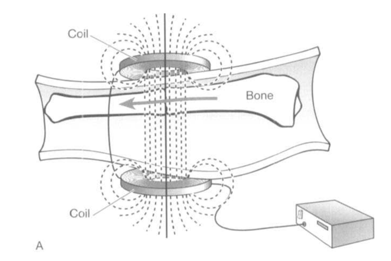
FDA Approved BONE Stimulator
This device generates a non-ionizing pulsed electromagnetic field with an intensity of approximately 2 Gauss and frequency components in the 1Hz-50KHz range. This field is distributed within and near the treatment coil.
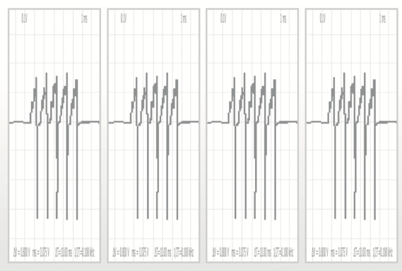
The dynamic characteristic of the field was well illustrated by a sequence of field isosurfaces corresponding to the evolution with time of the electric current waveform (sawtooth) powering the coils.
This device generates a non-ionizing pulsed electromagnetic field with an intensity of approximately 2 Gauss and frequency components in the 1Hz-50KHz range. This field is distributed within and near the treatment coil.
The dynamic characteristic of the field was well illustrated by a sequence of field isosurfaces corresponding to the evolution with time of the electric current waveform (sawtooth) powering the coils.
Safe and noninvasive solution that helps to promote healing in fractured bones and spinal fusions that have not healed or have difficulty healing.
PEMF stimulates the bones natural healing process by sending low level pulses of electromagnetic energy of PEMF to the injury or fusion sites.
PEMF has been shown to activate the body's natural healing process by acting at a molecular, cellular, and tissue level and has been clinically proven to improve bone healing rates of nonunion fracture or fusion.
Molecular Level
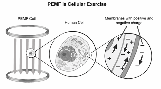
Exposure of a bone forming cell to PEMF generates an electrical field across the cell membrane.
*Panagopoulos DJ, Karabarbounis A, Margaritis LH
Mechanism for action of electromagnetic fields on cells
Biochem Biophys Res Commun 2002; 298: 95-102
PEMF stimulates the bones natural healing process by sending low level pulses of electromagnetic energy of PEMF to the injury or fusion sites.
PEMF has been shown to activate the body's natural healing process by acting at a molecular, cellular, and tissue level and has been clinically proven to improve bone healing rates of nonunion fracture or fusion.
Molecular Level
Exposure of a bone forming cell to PEMF generates an electrical field across the cell membrane.
*Panagopoulos DJ, Karabarbounis A, Margaritis LH
Mechanism for action of electromagnetic fields on cells
Biochem Biophys Res Commun 2002; 298: 95-102
Bone Cells recognize PEMF activating several pathways - leading to an immediate cellular response. (SIMULATES EXERCISE - LOAD/STRESS PIEZOELECTRIC.
Also anti-inflammatory molecules, NO, ATP, Calcium apatite crystals,
And collagen.
*Schnoke M, Midura RJ. Pulsed Electromagnetic fields rapidly modulate intracellular signaling events in osteoblastic cells: comparison to parathyroid hormone and insulin. J Orthop Res. 2007; 25(7): 933-40
*Vincenzi F, Targa M, Corciulo C, Gessi S, Merighi S, Setti S, Cadossi R, Goldring MB, Borea PA, Varani K. Pulsed Electromagnetic Fields increased the Anti-Inflammatory Effect of A2a and A3 Adenosine receptors in Human T/C-28a2 Chondrocytes and fFOB 1.19 Osteoblasts. PLOS ONE 2013; 8(5):e65561
Cellular Level
At the cellular level, the PEMF signal activates bone growth signaling pathways, which enhances bone formation by increasing both the population - cellular proliferation - and maturity of the bone building cells.
Osteoclasts, Osteoblasts, Chondroblasts, fibroblasts.
*Selvamurugan N, Kwok S, Vasilov A, Jefcoat SC, Patridge NC. Effects of BMP-2 and pulsed electromagnetic field (PEMF) on rat primary osteoblastic cell proliferation and gene expression. J Orthop Res. 2007; 25(9): 1213-20
Tissue Level
The exposure of the bone building cells enables an increase in the strength and quality of bone at the tissue level. Bone and Collagen.
*Selvamurugan N, He Z, Rifkin D, Dabovic B, Patridge NC. Pulsed Electromagnetic Field Regulates MicroRNA 21 Expression to Activate TGF-B Signaling in Human Bone Marrow Stroma Cells to Enhance Osteoblast Differentiation. Stem cells international, 2017 Apr 23; 2017.
Published pre-clinical research on fracture healing shows that the group treated with PEMF had a 2-fold increase in bone volume 13-20 days post fracture!!
Following PEMF exposure, the resulting changes to the molecular, cellular and tissue processes have been clinically shown to improve the healing rates of non-union fractures and spinal fusions.
* Ibiwaye MO, Powell Ka, Grabnier MD. Bone mass is preserved in a critical-sized osteotomy by low energy pulsed electromagnetic fields as quantitated by in vivo micro-computed tomography. J Orthop Res. 2004; 2004 22(5): 1086-93.
* Midura RJ, Ibawaye MO, Powell KA. Pulsed electromagnetic field treatments enhance the healing of fibular osteotomies. J Orthop Res. 2005; 23:1035-46
* Androjna C, Waldorff EJ, Ryaby JT, Zborowski M, Midura RJ. Optimizing Pulsed Electromagnetic Field (PEMF) Signals to Reduce Bone Loss Associated with Osteoporosis". Orthopedic Research Society March 2017, San Diego Calfornia.
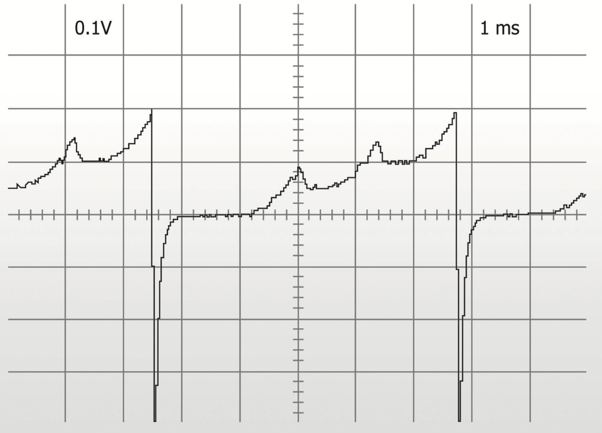
The sawtooth waveform - The KEY to induction
The most well-known signal shape is the sawtooth waveform introduced by Bassett in 1974. Dr. Bassett observed that changes in the electromagnetic signal induce
an electrical current within the treated tissue, with maximum current being induced when the signal changes most abruptly, namely when it falls from its peak value to its lowest value (fall time). The piezoelectric current induced within bone accelerated the bone healing.
|
|
Tell us about NASA’s research into PEMF?
NASA recognized a critical need to develop effective prevention and treatments for bone loss and muscle atrophy to enable future human space explorations on to the Moon, Mars and beyond. Bone loss causes increased risks of bone fracture and kidney stones, which can compromise astronaut health and mission objectives.
Consequently NASA mobilized its resources to develop methods that can enhance bone retention along with other benefits.
**Goodwin, T. Physiologic and molecular genetic effects of time-varying electromagnetic fields on human neuronal cells. Lyndon B Johnson Space Center. 2003 Sep. NASA
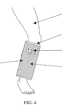
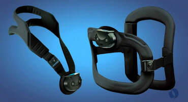
PEMFs were found to help reverse the effects of low gravity. The long term goal was to produce garments incorporating PEMF devices that could be worn by astronauts. Eventually NASA patented systems for this purpose. The patent claims that the apparatus is for enhancing tissue repair in mammals.
The apparatus shown here includes a sleeve with a PEMF coil that uses a SQUARE wave PEMF at VERY low intensity ~.05-.5 Gauss. Regeneration was found to be accelerated with the device.
**Goodwin TJ, Parker CR. 2007. Apparatus for enhancing tissue repair in mammals. U.S. Patent #7,179,217
The apparatus shown here includes a sleeve with a PEMF coil that uses a SQUARE wave PEMF at VERY low intensity ~.05-.5 Gauss. Regeneration was found to be accelerated with the device.
**Goodwin TJ, Parker CR. 2007. Apparatus for enhancing tissue repair in mammals. U.S. Patent #7,179,217
PEMF Therapy on Bones essentially works through Faraday's Law and the piezoelectric effect.
Bone production, whether in a person with osteoporosis or a non-healing fracture or having undergone spinal fusion surgery, can be stimulated by pulsating electromagnetic field exposure.
Possible mechanisms of action include
1) Faradays Law,
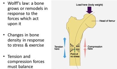
2) the Reverse piezoelectric effect which induces an electrical current within bone similar to the current produced by the natural tensile, bending and pressure forces on the bone; ===> Wolfs Law
3) by direct stimulation of osteoblasts and chondrocytes;
4) and by the removal of the bone-growth inhibition effect of parathyroid hormone.
What is so wonderful about using PEMF therapy or changing magnetic fields is that they penetrate deeper and work faster than electric currents AND they are safer and less invasive. Literally PEMF is 3D microcurrent therapy!
Through Faradays law PEMF therapy induces electric currents or charge in tissues, which improves the permeability of the cell membrane, increases cellular voltage and ATP which then increases bone formation and bone density!
Bone production, whether in a person with osteoporosis or a non-healing fracture or having undergone spinal fusion surgery, can be stimulated by pulsating electromagnetic field exposure.
Possible mechanisms of action include
1) Faradays Law,
2) the Reverse piezoelectric effect which induces an electrical current within bone similar to the current produced by the natural tensile, bending and pressure forces on the bone; ===> Wolfs Law
3) by direct stimulation of osteoblasts and chondrocytes;
4) and by the removal of the bone-growth inhibition effect of parathyroid hormone.
What is so wonderful about using PEMF therapy or changing magnetic fields is that they penetrate deeper and work faster than electric currents AND they are safer and less invasive. Literally PEMF is 3D microcurrent therapy!
Through Faradays law PEMF therapy induces electric currents or charge in tissues, which improves the permeability of the cell membrane, increases cellular voltage and ATP which then increases bone formation and bone density!
1.1. Piezoelectric effect - Pressure Electricity
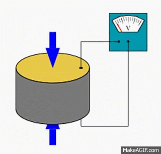
The word “piezo” is Greek and means “to press” (peizein = press or pressure). The prefix is used to describe the electrical charge of crystals or bone substances in response to mechanical pressure (stretching or squeezing). Or more accurately, piezoelectricity is the ability of certain crystals to produce a voltage when subjected to mechanical stress (the substance is squeezed or stretched.
The BONE IS CRYSTALLINE AND EXHIBITS PIEZOELECTRIC PROPERTIES!!
1. Piezoelectric effect and osteoblast proliferation
Previously one had assumed that the piezoelectric effect on its own was responsible for bone growth. Today, studies with bone cells have shown a direct stimulation with bone cells. To explain: With the help of the piezoelectric effect, one “simulates” the natural load on the bone. The bone receives growth signals that are transmitted to the bone formation cells.
If we put a compressive load on a bone the pressure is transmitted to the membrane of the bone cells. The result is the development of an electrical current within the bone that causes the bone to strengthen. (Hayashi 1998/74) This bone compression effect is generated by muscle tension across long bones with normal muscle contractions, by the Earth’s gravitational field, whole body vibration and by pulsating magnetic fields.
***Hayashi Y. Nippon Rinsho 1998 Jun; 56 (6): 1551-6.
Magnetic fields simulate the piezoelectric currents developed in bone by natural movement. (Dealer 1981/75) These currents cause cartilage and bone cells to proliferate, be renewed, or be regenerated together with the increase of appropriate messenger substances (cAMP, calcium ions, etc.). It is most impressive to witness the healing of complicated fractures evoked by magnetic fields when other healing approaches, including surgical fixation with screws and plates, have failed.
**Dealer SF. J Med Eng Technol 1981 Mar; 5 (2): 73-9.
Technically PEMFs and electrotherapy uses the "reverse piezoelectric effect" whereby the application of an electrical field creates mechanical deformation in the crystal or Vibrations with PEMFs as they are changing fields. PEMFs create circulating electric fields through Faraday's law.
**Wolfs law - The form of the bone elements place or displace themselves in the direction of the functional pressure and increase or decrease their mass to reflect the amount of functional pressure.
Use Strength Training, whole body vibration and Exercise in general to strenghten the bones via piezoelectric effect
AND PEMF the strengthen bones via CONVERSE Piezoelectric effect.
In My opinion the BEST OF THE BEST NATURAL APPROACH FOR BONE DENSITY IS
1) PEMF
2) Whole Body Vibration and strength training (and avoid sitting too much)
3) Bone Building Diet and Supplements
PEMF provides the energy or power and good nutrition and supplements provides the bricks and mortar to build new bone. PEMF both improves assimilation and absorption of the food and supplements AND the energy from PEMF helps to create new bone structures.
PEMFs repair bone and enhance bone tissue formation, through the enhancement of the natural formation and deposition of calcium phosphate crystal seeds in the bone (1).
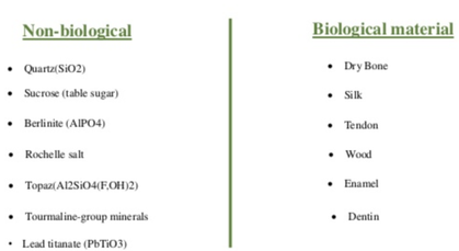
Crystalline Arrangements are the rule and not the exception in living systems. So MUSCLES, TENDONS, Bones, myelin, muscle, sensory organs and even cell membrane crystalline and therefore piezoelectric properties.
Virtually all the tissues in the body produce an electric field when they are stretched or compressed = ENERGY! These oscillating fields correspond precisely to the input stressors which mean they contain the information. This information is electrically and electronically conducted through the living matrix.
The electric fields produced during movements are widely considered to provide the information that directs the activities of generative cells (Bassett). These osteoblasts, myoblasts, fibroblasts and other 'stem' cells help to reform and heal tissues so the body can adapt to ways the body is used.
The mechanisms by which cells lay down or reabsorb materials in bone... Electric fields generated during movement (streaming or piezoelectric potentials) or PEMF signal cells (fibroblasts in connective tissue, osteoblasts in bones) to lay down collagen in the direction of tension/stress and therefore strengthen the tissue. With less loading or movement, the electric fields are weaker and less frequent, and the cells resorb collagen (Bassett 1968).
Related process are involved in wound healing... again electric fields signal.
Injuries create tension/stress in bone/tissues which generates electricity (piezoelectric effect and streaming potentials) which signals cells to migrate (by cytoskeleton motors that consume chemical energy, and then cells lay down collagen!
3) PEMF Increases Stem Cells Energetically
PEMF Increases Stem Cell Production and Differentiation
**Beckers Study and Cell De-differentiation with Cellular Voltage
There is a significant amount of evidence that PEMFs affect stem cells.
PEMF stimulation increases the amount of viable stem cells by 40-59% and results in up to 60% higher cell densities. PEMF has also been proven and shown to help stem cells differentiate which is crucial for healing and regeneration, for example differentiating neural stems cells into neurons.
Li Y, Zhoa L, Xing X. Effects of different frequency electromagnetic fields on the differentiation of midbrain neural stems cells. Space Med Eng (Beijing). 2002 Oct; 15(5): 374-6.
NASA studied the use of 10 Hz low intensity square wave PEMF stimulation on the growth of nerve stem cells. With this particular 10 Hz signal, NASA discovered about a 400% increases in neural stem cells and the signal turned on about 160 growth and regeneration genes.
Goodwin, T. Physiologic and molecular genetic effects of time-varying electromagnetic fields on human neuronal cells. Lyndon B Johnson Space Center. 2003 Sep. NASA
4) Inhibit Bone-Growth Inhibiting Effects of Parathyroid
Physical stresses such as compression or shear forces, movements, hormones and enzymes stimulate bone growth and repair. Pulsating magnetic fields represent a profoundly powerful signal for the stimulation of bone growth. In a magnetic field, bone cells lose their sensitivity to the bone-growth inhibiting effects of parathyroid hormone, so osteoblasts are unopposed and unhindered in their bone-building activities. Many studies, including research at the Institut für Strahlenforschung München (Institute for Radiation Research) in Munich, have shown that osteoblasts exposed to a biologically active magnetic field form significantly more Type 1 colla- gen than unexposed control cells. (Indouraine 2001/76, Guldner 1999/77, Heermeier 1998/78).
Physical stresses such as compression or shear forces, movements, hormones and enzymes stimulate bone growth and repair. Pulsating magnetic fields represent a profoundly powerful signal for the stimulation of bone growth. In a magnetic field, bone cells lose their sensitivity to the bone-growth inhibiting effects of parathyroid hormone, so osteoblasts are unopposed and unhindered in their bone-building activities. Many studies, including research at the Institut für Strahlenforschung München (Institute for Radiation Research) in Munich, have shown that osteoblasts exposed to a biologically active magnetic field form significantly more Type 1 colla- gen than unexposed control cells. (Indouraine 2001/76, Guldner 1999/77, Heermeier 1998/78).
5) PEMF Nitric Oxide
Connective Tissue - PEMF Helps Regenerate ALL types of Connective Tissue
Suffixes -blast, -clasts and cytes indicate functions in specialized cells in the connective tissue
-Blasts create extracellular matrix
-Cytes Maintain extracellular matrix
-Clasts destroy extracellular matrix
In hundreds of research studies (including those leading to FDA approval) PEMFs are seen to enhance the proliferation and maturation of bone building cells (osteoblasts) and thus the formation of new bone. To rebuild bone in a fracture also requires the OLD bone to be broken down and removed. The cells that breakdown bone are call osteoclasts and are ALSO increase by PEMFs.
So PEMFs not only stimulate the bone building molecules (osteoblasts) but also help with the remodeling molecules (osteoclasts).
These effects are as good or better and safer than medications used for this purpose. And typically medications do not affect BOTH functions (blasts and clasts). As result, medications often leave the bone even weaker in the long run.
|
|
Blood Vessels in the Bones are Very Complex - So Much so, just last year, new discoveries were made!!
https://www.sbbs-soc.com/2019/01/30/scientists-discover-new-blood-vessels-in-bone/
”It’s totally crazy there are still things to find out about human anatomy – we have discovered blood vessels in a new place that we didn’t know about before.”
Matthias Gunzer, Study Author
5) Nitric Oxide - KEYS TO Strong Bones from PEMF
Energy, Nitric Oxide, Inflammation, DNA Calcium Absorption (microcirculation).
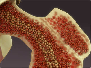
Bones are not hard and Dry , they are RICH with Blood Vessels.
PEMF improves Inflammation and the Production of NO. Inflammation is known to hinder bone repair, NO known to improve it!
One of the ways PEMF helps with Strong bones is increased microcirculation via enhanced Nitric Oxide (NO). Many studies show NO is an essential part of healing bones and enhancing the very complicated blood vessel networks in the bones.
Studies on bone proliferation have proven that nitric oxide synthesis is increased by exposure to pulsating magnetic fields. (Diniz 2002/102)
Low frequency PEMFs change Calcium Ion Concentrations in cells and stimulate DNA production. The calcium Ions inside cells regulate NO production and increase it. NO in bone cells has been linked to the development and growth into mature bone cells.
These effects of PEMFs are important for the management of osteopenia or osteoporosis, fractures and other bone problems!!
https://www.sbbs-soc.com/2019/01/30/scientists-discover-new-blood-vessels-in-bone/
”It’s totally crazy there are still things to find out about human anatomy – we have discovered blood vessels in a new place that we didn’t know about before.”
Matthias Gunzer, Study Author
5) Nitric Oxide - KEYS TO Strong Bones from PEMF
Energy, Nitric Oxide, Inflammation, DNA Calcium Absorption (microcirculation).
Bones are not hard and Dry , they are RICH with Blood Vessels.
PEMF improves Inflammation and the Production of NO. Inflammation is known to hinder bone repair, NO known to improve it!
One of the ways PEMF helps with Strong bones is increased microcirculation via enhanced Nitric Oxide (NO). Many studies show NO is an essential part of healing bones and enhancing the very complicated blood vessel networks in the bones.
Studies on bone proliferation have proven that nitric oxide synthesis is increased by exposure to pulsating magnetic fields. (Diniz 2002/102)
Low frequency PEMFs change Calcium Ion Concentrations in cells and stimulate DNA production. The calcium Ions inside cells regulate NO production and increase it. NO in bone cells has been linked to the development and growth into mature bone cells.
These effects of PEMFs are important for the management of osteopenia or osteoporosis, fractures and other bone problems!!
Osteoporosis - A Silent Killer
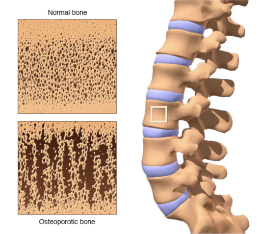
Also called "brittle bone disease," osteoporosis makes bone more fragile and increases your chance of sustaining a broken bone. Bone has a lattice-shaped structure, much like a honeycomb.
Osteoporosis is defined as a Skeletal Disorder, a disorder of the bones in which there is a reduction in the amount of strength in the bones, the bones are less strong than they should be because of in most cases a loss of bone which occurs as we get older.
And accompanying that loss of bone is a damage to the bone structure and the bones become weakened by this and as a consequence are much more likely to break.
So with osteoporosis what we are trying to do is prevent broken bones.
Many people have no symptoms until they have a bone fracture.
Four Important Things to Know About Osteoporosis
1) Osteoporosis is common; about 10 million Americans have it Osteoporosis is most common in women, as there is accelerated loss of bone following menopause. The two most critical factors in determining who gets osteoporosis are how much bone mass an individual accumulates in their teens and twenties, and how quickly they lose it thereafter.
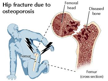
2) Half of women over age 50 will sustain a broken bone resulting from osteoporosis. The major complication of osteoporosis is a fractured bone. Many fractures resulting from osteoporosis can have major health implications. Spine and hip fractures are notorious for leading to significant declines in function and overall health.
3) After the age of 30, you lose bone rather than gain it. That said, there are steps that you can take to slow the rate of bone loss. This is why bone health in young people, particularly young women, is so critical. If they don't build bone in their teenage years, they will have a much higher change of developing osteoporosis later in life.
4) There are ways to control osteoporosis. While some aspects that determine bone density are of of your control (race, gender, etc.), there are others that you can influence (diet, exercise, etc.) Studies show that factors you can't control account for 75 percent of the condition, but the other 25 percent is up to you.
Risk Factors
Female gender
Caucasian race
Advanced age
Slender build or fair skin
Poor nutrition
Tobacco use
Some specific medications (e.g. steroids)
Some medical conditions (e.g. thyroid abnormalities)
Broken bones are the often the end result of osteoporosis. The goal of treatment is to prevent sustaining a broken bone, especially a broken hip. Some of the more common fractures that occur as a result of osteoporosis include:
*Hip fractures
*Spine compression fractures
Wrist fractures
Shoulder fractures
Pelvis fractures
Tibial plateau fractures
Ankle fractures
Also called "brittle bone disease," osteoporosis makes bone more fragile and increases your chance of sustaining a broken bone. Bone has a lattice-shaped structure, much like a honeycomb.
Osteoporosis is defined as a Skeletal Disorder, a disorder of the bones in which there is a reduction in the amount of strength in the bones, the bones are less strong than they should be because of in most cases a loss of bone which occurs as we get older.
And accompanying that loss of bone is a damage to the bone structure and the bones become weakened by this and as a consequence are much more likely to break.
So with osteoporosis what we are trying to do is prevent broken bones.
Many people have no symptoms until they have a bone fracture.
Four Important Things to Know About Osteoporosis
1) Osteoporosis is common; about 10 million Americans have it Osteoporosis is most common in women, as there is accelerated loss of bone following menopause. The two most critical factors in determining who gets osteoporosis are how much bone mass an individual accumulates in their teens and twenties, and how quickly they lose it thereafter.
2) Half of women over age 50 will sustain a broken bone resulting from osteoporosis. The major complication of osteoporosis is a fractured bone. Many fractures resulting from osteoporosis can have major health implications. Spine and hip fractures are notorious for leading to significant declines in function and overall health.
3) After the age of 30, you lose bone rather than gain it. That said, there are steps that you can take to slow the rate of bone loss. This is why bone health in young people, particularly young women, is so critical. If they don't build bone in their teenage years, they will have a much higher change of developing osteoporosis later in life.
4) There are ways to control osteoporosis. While some aspects that determine bone density are of of your control (race, gender, etc.), there are others that you can influence (diet, exercise, etc.) Studies show that factors you can't control account for 75 percent of the condition, but the other 25 percent is up to you.
Risk Factors
Female gender
Caucasian race
Advanced age
Slender build or fair skin
Poor nutrition
Tobacco use
Some specific medications (e.g. steroids)
Some medical conditions (e.g. thyroid abnormalities)
Broken bones are the often the end result of osteoporosis. The goal of treatment is to prevent sustaining a broken bone, especially a broken hip. Some of the more common fractures that occur as a result of osteoporosis include:
*Hip fractures
*Spine compression fractures
Wrist fractures
Shoulder fractures
Pelvis fractures
Tibial plateau fractures
Ankle fractures
|
|
|
|
What is Osteoporosis and What Causes It?
Osteoporosis is a bone disease that occurs when the body loses too much bone, makes too little bone, or both. As a result, bones become weak and may break from a fall or, in serious cases, from sneezing or minor bumps.
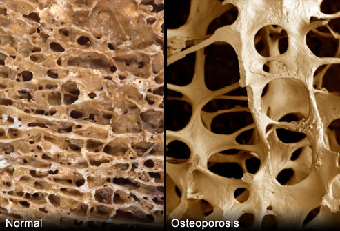
Osteoporosis means “porous bone.” Viewed under a microscope, healthy bone looks like a honeycomb. When osteoporosis occurs, the holes and spaces in the honeycomb are much larger than in healthy bone. Osteoporotic bones have lost density or mass and contain abnormal tissue structure. As bones become less dense, they weaken and are more likely to break.
Osteoporosis is Common
About 54 million Americans have osteoporosis and low bone mass, placing them at increased risk for osteoporosis. Studies suggest that approximately one in two women and up to one in four men age 50 and older will break a bone due to osteoporosis.
Osteoporosis is Serious
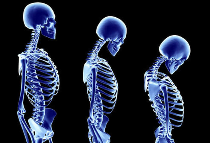
Breaking a bone is a serious complication of osteoporosis, especially with older patients. Osteoporotic bone breaks are most likely to occur in the hip, spine or wrist, but other bones can break too. In addition to causing permanent pain, osteoporosis causes some patients to lose height. When osteoporosis affects vertebrae, or the bones of the spine, it often leads to a stooped or hunched posture.
Osteoporosis may limit mobility, which often leads to feelings of isolation or depression. Additionally, twenty percent of seniors who break a hip die within one year from either complications related to the broken bone itself or the surgery to repair it. Many patients require long-term nursing home care.
Osteoporosis Can Sneak up on You
Osteoporosis is often called a silent disease because one can’t feel bones weakening. Breaking a bone is often the first sign of osteoporosis or a patient may notice that he or she is getting shorter or their upper back is curving forward. If you are experiencing height loss or your spine is curving, be sure to consult your doctor or healthcare professional immediately.
Osteoporosis is a bone disease that occurs when the body loses too much bone, makes too little bone, or both. As a result, bones become weak and may break from a fall or, in serious cases, from sneezing or minor bumps.
Osteoporosis means “porous bone.” Viewed under a microscope, healthy bone looks like a honeycomb. When osteoporosis occurs, the holes and spaces in the honeycomb are much larger than in healthy bone. Osteoporotic bones have lost density or mass and contain abnormal tissue structure. As bones become less dense, they weaken and are more likely to break.
Osteoporosis is Common
About 54 million Americans have osteoporosis and low bone mass, placing them at increased risk for osteoporosis. Studies suggest that approximately one in two women and up to one in four men age 50 and older will break a bone due to osteoporosis.
Osteoporosis is Serious
Breaking a bone is a serious complication of osteoporosis, especially with older patients. Osteoporotic bone breaks are most likely to occur in the hip, spine or wrist, but other bones can break too. In addition to causing permanent pain, osteoporosis causes some patients to lose height. When osteoporosis affects vertebrae, or the bones of the spine, it often leads to a stooped or hunched posture.
Osteoporosis may limit mobility, which often leads to feelings of isolation or depression. Additionally, twenty percent of seniors who break a hip die within one year from either complications related to the broken bone itself or the surgery to repair it. Many patients require long-term nursing home care.
Osteoporosis Can Sneak up on You
Osteoporosis is often called a silent disease because one can’t feel bones weakening. Breaking a bone is often the first sign of osteoporosis or a patient may notice that he or she is getting shorter or their upper back is curving forward. If you are experiencing height loss or your spine is curving, be sure to consult your doctor or healthcare professional immediately.
By Understanding How PEMF heals the BONES like fractures, breaks, we can understand not only how PEMF then helps with Osteoporosis BUT healing ALL types of connective tissue!
In order to adequately perform its functions as a skeletal framework, rigid protector of the spinal column and attachment point for muscles, tendons and ligaments, bones must have a great deal of tensile strength, compression strength, and elasticity. In order to fulfill these tasks, the bone consists of a thick, solid outer layer called the cortex, and a spongy interior with tiny rigid crossbeams called trabeculae.
The dual cortical-spongy bone structure is comparable to steel-and-concrete construction. The “steel supports” are equivalent to the cortical bone beams and the collagen fibers used to form them. The “concrete” is the pressure resistant bone minerals (calcium and phosphate) within the spongy bone.
Bone is a dynamic, living tissue. This fact can be lost by the fact that all models of bone are dry. We begin to view bone as a dry, inert substance, like wood. However, in the living body bone is a reservoir of calcium, a factory for producing blood cells, an attachment site for contraction, a hinge-point for joint movement, and even a delicate vibratory sensor, as in the tiny bones of the inner ear – the incus, stapes and malleus.
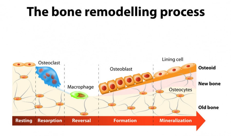
Bone matrix (or “osteoid”) undergoes continuous breakdown and rebuilding. Osteoblasts are bone-forming cells. They incorporate calcium and other minerals into living bone matrix. Osteoblasts build mature bone cells. Osteoclasts remove bone cells. They break down bone substance. Osteoclasts prevent the formation of too much bone. Since bone is a living tissue, it has a “turnover rate.” Old cells are replaced with new ones. The osteoblasts and osteoclasts must operate in a balanced fashion for this turnover to occur normally. In healthy bone there is equilibrium between the building forces (osteoblasts) and the breaking down forces (osteoclasts). The bones we have today will have been completely renewed.
Osteoblasts produce the bone substance. In addition to calcium and phosphate, they also use collagen to give the bone its extra- ordinary tensile strength. This is contrasted to eggshells which are also made of calcium but which have considerably less strength and more fragility than living bone.
Physical stresses such as compression or shear forces, movements, hormones and enzymes stimulate bone growth and repair. Pulsating magnetic fields represent a profoundly powerful signal for the stimulation of bone growth. In a magnetic field, bone cells lose their sensitivity to the bone-growth inhibiting effects of parathyroid hormone, so osteoblasts are unopposed and unhindered in their bone-building activities. Many studies, including research at the Institut für Strahlenforschung München (Institute for Radiation Research) in Munich, have shown that osteoblasts exposed to a biologically active magnetic field form significantly more Type 1 colla- gen than unexposed control cells. (Indouraine 2001/76, Guldner 1999/77, Heermeier 1998/78).
Summary/effect:
Bone production, whether in a person with osteoporosis or a non-healing fracture or having undergone spinal fusion surgery, can be stimulated by pulsating electromagnetic field exposure. Possible mechanisms of action include the piezoelectric effect, which induces an electrical current within bone similar to the current produced by the natural tensile, bending and pressure forces on the bone; by direct stimulation of osteoblasts and chondrocytes; and by the removal of the bone-growth inhibition effect of parathyroid hormone. Other mechanisms have recently been elucidated and are discussed later in this text.
In order to adequately perform its functions as a skeletal framework, rigid protector of the spinal column and attachment point for muscles, tendons and ligaments, bones must have a great deal of tensile strength, compression strength, and elasticity. In order to fulfill these tasks, the bone consists of a thick, solid outer layer called the cortex, and a spongy interior with tiny rigid crossbeams called trabeculae.
The dual cortical-spongy bone structure is comparable to steel-and-concrete construction. The “steel supports” are equivalent to the cortical bone beams and the collagen fibers used to form them. The “concrete” is the pressure resistant bone minerals (calcium and phosphate) within the spongy bone.
Bone is a dynamic, living tissue. This fact can be lost by the fact that all models of bone are dry. We begin to view bone as a dry, inert substance, like wood. However, in the living body bone is a reservoir of calcium, a factory for producing blood cells, an attachment site for contraction, a hinge-point for joint movement, and even a delicate vibratory sensor, as in the tiny bones of the inner ear – the incus, stapes and malleus.
Bone matrix (or “osteoid”) undergoes continuous breakdown and rebuilding. Osteoblasts are bone-forming cells. They incorporate calcium and other minerals into living bone matrix. Osteoblasts build mature bone cells. Osteoclasts remove bone cells. They break down bone substance. Osteoclasts prevent the formation of too much bone. Since bone is a living tissue, it has a “turnover rate.” Old cells are replaced with new ones. The osteoblasts and osteoclasts must operate in a balanced fashion for this turnover to occur normally. In healthy bone there is equilibrium between the building forces (osteoblasts) and the breaking down forces (osteoclasts). The bones we have today will have been completely renewed.
Osteoblasts produce the bone substance. In addition to calcium and phosphate, they also use collagen to give the bone its extra- ordinary tensile strength. This is contrasted to eggshells which are also made of calcium but which have considerably less strength and more fragility than living bone.
Physical stresses such as compression or shear forces, movements, hormones and enzymes stimulate bone growth and repair. Pulsating magnetic fields represent a profoundly powerful signal for the stimulation of bone growth. In a magnetic field, bone cells lose their sensitivity to the bone-growth inhibiting effects of parathyroid hormone, so osteoblasts are unopposed and unhindered in their bone-building activities. Many studies, including research at the Institut für Strahlenforschung München (Institute for Radiation Research) in Munich, have shown that osteoblasts exposed to a biologically active magnetic field form significantly more Type 1 colla- gen than unexposed control cells. (Indouraine 2001/76, Guldner 1999/77, Heermeier 1998/78).
Summary/effect:
Bone production, whether in a person with osteoporosis or a non-healing fracture or having undergone spinal fusion surgery, can be stimulated by pulsating electromagnetic field exposure. Possible mechanisms of action include the piezoelectric effect, which induces an electrical current within bone similar to the current produced by the natural tensile, bending and pressure forces on the bone; by direct stimulation of osteoblasts and chondrocytes; and by the removal of the bone-growth inhibition effect of parathyroid hormone. Other mechanisms have recently been elucidated and are discussed later in this text.
1. Treatment of bones, non-healing (nonunion) fractures, spine fusions and non-healing wounds
General information:
Prior to 1993, 250,000 patients in the United States had been treated successfully for bone and wound healing disorders using pulsating magnetic fields. In the USA medical applications of pulsating magnetic field therapy occupy a small sector in orthopedic medicine and endocrinology that deals with bone healing, improving spinal fusion rates, and the healing of ulcers from diabetes, venous stasis, and pressure sores in bed-ridden or wheelchair-ridden people. Pulsating magnetic fields are FDA registered (since 1979) and are marketed to the orthopedic profession in the United States as “a bone stimulators.”13
A review of pulsating electromagnetic field (PEMF) therapy studies published in 2000 stated that, in the case of bone healing disorders, PEMFs are a “proven treatment with the potential for use for arthritis, bony necrosis, osteoporosis and wound healing disorders.” (Trock 2000/109)
Osteoporosis
According to the National Osteoporosis Foundation, about 85-90% of adult bone mass is acquired by age 18 in girls and age 20 in boys. Building strong bones during childhood can help prevent osteoporosis later in life. Based on the quality of the American diet, osteoporosis will predictably increase in the coming years. Of the 10 million Americans with osteoporosis, 8 million are women and 2 million are men. Costs associated with the treatment of osteoporosis are expected to increase from $19 billion in 2005 to over $25 billion by 2025. There has been a fivefold increase in office visits for osteoporosis (from 1.3-6.3 million) in the past 10 years.
Scientific literature on PEMFs and osteoporosis:
The non-dominant forearms of 20 osteoporosis-prone women were exposed to magnetic fields for 12 weeks. Pre- and post-treatment bone mineral density was measured using single-photon densitometry. Result: Significant and immediate increase in bone density during the 12-week treatment period.“ Cross-over” increases in bone density were also noted in the opposite, untreated arm. At 36 weeks (24 weeks after treatment ceased), the density of the bone began to decrease slightly. (Tabrah 1990) An 8-year follow-up measurement of the same group of women showed maintenance of the increased bone density over time. (Tabrah 1998/110)
Another study examined the effect of PEMFs on bone formation and disuse osteoporosis sustained during limb lengthening in a double-blind study. Seven males and six females were randomly allocated to receive either an active or an inactive PEMF coil. Result: Stimulation with PEMFs was shown to prevent bone loss adjacent to the distraction gap following limb-lengthening surgery. Bone loss in the sham treatment group was 54%, but in the active treatment group, only 10% at 12 months post-surgery. (Eyres 1996/115)
Osteporosis
Increases risks to hip and spine fractures that can be crippling.
PEMFs penetrate bone without any blockage or absorption, meaning PEMF can stimulate the WHOLE volume of BONE unlike any other approach in energy medicine.
PEMF provides the energy or power and good nutrition and supplements provides the bricks and mortar to build new bone. PEMF both improves assimilation and absorption of the food and supplements AND the energy from PEMF helps to create new bone structures.
PEMFs repair bone and enhance bone tissue formation, through the enhancement of the natural formation and deposition of calcium phosphate crystal seeds in the bone (1).
Catch it at Osteopenia and it will be much easier to reverse, but even with Osteoporosis good results have been seen with PEMF + Nutrition vs high risk drugs (though it can take time).
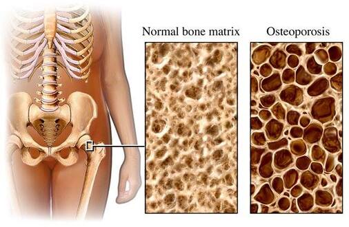
Description
Osteoporosis means “porous bones.” Due to low levels of calcium, phosphorus and other minerals the bones become weak and brittle. Fractures can occur with minor exertions such as lifting a grocery bag, bending over, or even coughing.
Bone demineralization is the cause of osteoporosis. This occurs when bone minerals are lost faster than they are replaced. Peak bone mass is achieved around the age of 35. After this, there are slight losses in bone density each year. At menopause, when estrogen levels drop, bone loss can increase dramatically. Osteopenia refers to mild bone loss that isn’t severe enough to be called osteoporosis, but that increases the risk of osteoporosis. Doctors can detect osteopenia or early signs of osteoporosis using a variety of devices to measure bone density.
In addition to a drop in estrogen levels with menopause, other risk factors associated with the development of osteoporosis include gender, age, race (increased risk when one is white or from Southeast Asia), family history, frame size, tobacco use, eating disorders, corticosteroid medications, hyperthyroidism, the use of medications (including methotrexate, SSRIs, diuretics, anti-seizure medications and antacids), medical conditions that decrease calcium absorption, breast cancer, low calcium intake, sedentary lifestyle, excessive soda consumption, chronic alcoholism, and depression.
A common result of osteoporosis is fractures. These usually occur in the spine, hip or wrist. One third of women over 65 will have vertebral fractures and 90% of women over the age of 75 have evidence of osteoporosis. Many people think osteoporosis is a disease of women only. However, it also affects men, and the prevalence of osteoporosis in men will increase in the coming decade.
A common result of osteoporosis is fractures. These usually occur in the spine, hip or wrist. One third of women over 65 will have vertebral fractures and 90% of women over the age of 75 have evidence of osteoporosis. Many people think osteoporosis is a disease of women only. However, it also affects men, and the prevalence of osteoporosis in men will increase in the coming decade.
According to the National Osteoporosis Foundation, there were over 2 million fractures related to osteoporosis in the United States in 2005. There were nearly 300,000 hip fractures, nearly 550,000 vertebral fractures, nearly 400,000 wrist fractures, and over 800,000 fractures at other sites combined. These fractures lead to an impaired quality of life from pain, vertebral compression, and loss of ambulation. Six months after a hip fracture, only 15% of people can walk across the room unaided.
Osteoporosis is a largely unrecognized potential cause of death. Osteoporosis-related fractures are often the precipitating event that leads to permanent infirmity and terminal decline. In 24% of those over 50 years of age suffering from hip fractures, death occurs within 12 months. A woman’s risk of osteoporosis-related hip fracture is equal to her risk of developing breast, uterine and ovarian cancer combined. Osteoporosis-related fractures can be expected to double during the next 5 decades.
In the early stages of bone loss, there are no symptoms. However, once the bone is weakened sufficiently, symptoms arise from fracture or from vertebral collapse. People with osteoporosis may observe loss of height over time, accompanied by a stooped posture called “kyphosis.”
Several factors can influence healthy levels of bone mineralization, and bone density. These factors include regular exercise, adequate amounts of dietary calcium and vitamin D, and good hormone balance. The effects of pulsating magnetic fields on bone density have been proven through extensive research. In animal studies, the use of pulsed electromagnetic fields was shown to be effective in restoring the trabecular bone structure in 35 Sprague- Dawley rats. (Chang 2003a/221; Chang 2003b/222) Rubin, McLeod et al induced osteoporosis in the ulna bone of turkeys, noting a 13% loss of bone compared to intact contralateral controls. By using a treatment regimen of one hour per day of pulsed electromagnetic fields, these researchers observed a bone-forming effect that they believed would be useful in treating patients with osteoporosis, those astronauts living for prolonged periods in weightless environments, and immobilized patients. (Rubin 1989/223)
In humans, researchers conducted a study with eight-year follow-up on changes to bone density after short-term electromagnetic field therapy. (Tabrah 1990) Using single photon absorptiometry to reliably measure changes in bone mineral density in a group of postmenopausal women with osteoporosis, they showed significantly increased bone density of the radius (a forearm bone commonly fractured at the wrist in these women) treated with pulsed magnetic fields. Eight years later, they showed maintenance of increased bone density of the treated radius bones over time, even though the therapy had been applied only for a short term in the first year of the experiment.
The scientific literature is replete with examples of restored bone density in cases of osteonecrosis of the hip, of non-healing fracture, and of up-regulated bone formation in a variety of circumstances. While the topic is somewhat exhaustive, it may be of interest, so the reader is invited to visit PubMed27 and, for example, perform an author search on “Bassett CA” (Dr. Andrew Bassett). Over 100 citations will be returned, many of them discussing PEMFs and bone metabolism.
The mechanisms of bone restoration using pulsed electromagnetic field therapy have been explored widely, and include the induction of piezoelectricity, desensitization of parathyroid hormone receptors, signaling of extracellular bone matrix and transmembrane proteins (to increase synthesis of bone proteins via the “mTOR” pathway and stimulation of osteoblastic proliferation and differentiation). (Patterson 2006/224)
Bone Building Action Plan
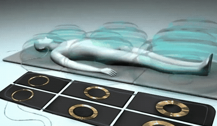
Settings and application
Use the whole body mat applicator three times per day for 8-16 minutes per application. Start at an intensity setting of 25 in the morning, and work toward 50 or 100, or to your preference. Use the mat at a setting of 50-100 at noon, and add 10-25 in the evening. Use the pad once daily for 24 minutes at the chest and lumbar spine areas at a setting of 100-150.
If you are experiencing spinal pain from osteoporosis, you may experience pain relief in as soon as 4-6 weeks. Long-lasting changes in bone density may be observed in 12-18 months or sooner. Repeat bone density testing at 12 months. Even if bone density is not increased in one year, the progression of the disease may be arrested by including the daily use of the MRS 2000 in your regimen of supportive treatments. Keep going! There may be a slight intensification of pain in approximately 15% of patients who experience pain from osteoporosis. This pain usually disappears within 7-10 days. Experience has shown 80% good to very good results with the use of the MRS 2000.
Nutritional Support for Bones
Protein: Did you know protein makes up about 50% of the volume of your bones, and roughly one-third of total bone mass?
Calcium: You’ve heard that calcium is a necessary mineral for your bones. But your body has hundreds of uses for calcium, like maintaining strong and healthy teeth, helping your muscles contract, letting your nerves send and receive signals, and much more. Yet only 32% of US adults are getting enough through their diet!2
Nuts/almonds, sesame seeds
Vitamin D3: One of vitamin D3’s major roles is to help your body efficiently utilize calcium.3 Vitamin D3 helps maintain normal blood levels of calcium and phosphorous and helps your body absorb calcium.
Because the best natural source of vitamin D3 is actually the sun. The sun’s UV rays trigger vitamin D production in your skin.4
Vitamin K2: There are several forms of vitamin K2. But one stands above the rest when it comes to healthy bones: K2-7. Vitamin K2-7 is crucial for calcium regulation. That’s because vitamin K2-7 acts as a calcium recycler! It transports excess calcium from soft tissues- like your arteries, kidneys, and eyes- and deposits it into your bones.
Vitamin C: Vitamin C promotes strong bones by increasing the solubility of calcium. That means vitamin C helps you absorb more calcium. What’s more, vitamin C also helps build the collagen base of your bones, giving them the supportive structure they need. So it’s important you’re getting adequate vitamin C to move out of osteoporosis!
The 12 minerals besides calcium you need are: magnesium, boron, copper, manganese, potassium, phosphorous, nickel, vanadium, zinc, silica, selenium, and strontium. Each of these minerals plays its own role in forming new bone.
For example, silica stimulates osteoblasts (your bone-building cells), and slows down osteoclasts (your bone-resorbing cells).5
Manganese stimulates bone-building enzymes, so much so in fact, that manganese helped increase bone density in postmenopausal women in one study.6 Another showed a manganese deficiency led to accelerated bone loss!7
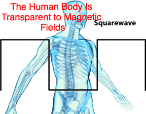
Body is Transparent to a Magnetic Field. Body does not reduce through body. Like wind blowing through a field of grass. Low Frequencies go all the way through. Long wavelengths PEMF Treats entire volume of body, not just surface.
PEMFs penetrate bone without any blockage or absorption, meaning PEMF can stimulate the WHOLE volume of BONE unlike any other approach in energy medicine.
PEMFs penetrate bone without any blockage or absorption, meaning PEMF can stimulate the WHOLE volume of BONE unlike any other approach in energy medicine.
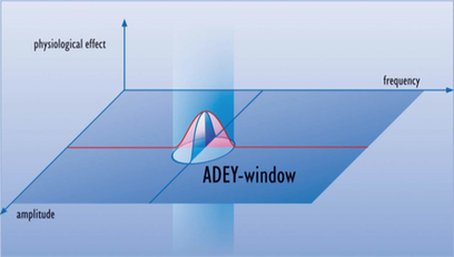
Biomedicial researchers have been testing the use of pulsing magnetic fields originating outside the body to induce microcurrents within tissues to stimulate healing.
A consistent observation is that triggering a cellular response requires the applicaton of energy in a very narrow range of frequencies and intensities. Extensive research on fracture non-unions using low intensity and frequency.
Microampere currents induced from outside the body restart the healing process by recruiting bone-forming cells in a manner similar to a natural repair response. Field effects are highly specific and confined to a narrow power frequency window. To high an induced current stimulates necrosis rather than repair! And the characteristics of a bone healing pulse are different from those of an osteonecrosis pulse. (Oschman Energy medicine pg 271).
Medical researchers have documented a cascade of signal transduction processes from the cell membrane to the nucleus and on to the genetic material that are facilitated by PEMF therapies (Bassett 1995). High intensity PEMF and high frequency electrosmog appear to have negative effects along the same pathways! (Oschman pf 271).
In General, organisms are poised to respond to minute 'whispers' in the electromagnetic environment. Bassett (1995) suggested an analogy between the arrays of bone cells in the osteon and the phased arrays of radio telescope antennas.
Radio telescope arrays enable astrophysicists to detect extremely weak electromagnetic signals from millions, even billions of light years away.
Bassett suggested that tissues extract information from fields originating outside the body by a process akin to that involved in radio astronomy.
These crystalline arrays are throughout the whole body so this applies NOT just to bone...
MORE IS NOT NECESSSARY!! The Body can tune to weak signals!!
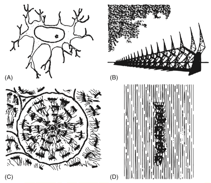
|
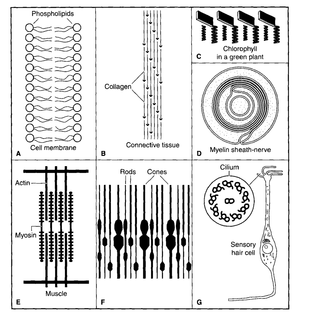
|
Further Proof MORE is not better
1) When the field strength is relatively high at 7 mT (milliTesla) osteoblasts were stimulated to increase bone production only at the beginning of the experiment. (Diniz 2002/81) Later in the study the osteoblast activity decreased. This is a reminder of the importance of the biological window – and in this case, the amplitude window in exerting consistently positive therapeutic effects. Most beneficial effects occur at very low electromagnetic amplitudes (or “flux densities”).
Diniz, P., K. Soejima, et al. (2002). Nitric oxide mediates the effects of pulsed electromagnetic field stimulation on
the osteoblast proliferation and differentiation. Nitric Oxide 7(1): 18-23.
2) Research shows that the induced field must be small or it will not work (Rubin 1989).
Rubin, C. T., K. J. McLeod, et al. (1989). Prevention of osteoporosis by pulsed electromagnetic fields. J Bone Joint Surg Am 71(3): 411-7.

Further Proof Low Intensity PEMF WORKS - 12 Studies!!
https://www.ncbi.nlm.nih.gov/pmc/articles/PMC3780531/
Very weak energy fields at the appropriate frequencies can be profoundly therapeutic. Other frequencies can produce pathological responses (like 4G).
While it may go against intuition, it appears that, within limits, it is not the strength of the signal that determines whether it will be beneficial or harmful, but rather it is the frequency!
Resonance is the reason for this frequency specificity. Biological effects like molecular resonances, are very frequency specific. This is vital for PEMF therapy along with all of energy healing and energy medicine devices.
For a long time scientists were very suspicious about the idea that very tiny energy fields could have any biological effects, let alone profound effects.
More and more evidence is proving that frequency resonance is the key to therapeutic energy healing. A recent study (Pall, 2013) points out that the answer to this question has been hiding in plain sight in the scientific literature for a long time.
An example of this is the relation to bone repair facilitated by exposure to tiny pulsating fields. The fact of this this therapeutic success in increasing osteoblast differentiation and maturation is difficult to challenge, because it is has been the subject of so many studies.
Pall lists these studies for the skeptical reader:
1. Ryabi JT. Clinical effects of electromagnetic fields on fracture healing. Clin Orthop Relat Res. 1998;355(Suppl. l):S205–15.
2. Oishi M, Onesti ST. Electrical bone graft stimulation for spinal fusion: a review. Neurosurgery. 2000;47:1041–55.
3. Aaron RK, Ciombor DM, Simon BJ. Treatment of nonunions with electric and electromagnetic fields. Clin Orthop Relat Res. 2004;10:579–93.
4. Goldstein C, Sprague S, Petrisor BA. Electrical stimulation for fracture healing: current evidence. J Orthop Trauma. 2010;24(Suppl. 1):S62–5.
5. Demitriou R, Babis GC. Biomaterial osseointegration enhancement with biophysical stimulation. J Musculoskelet Neuronal Interact. 2007;7:253–65.
6. Griffin XL, Warner F, Costa M. The role of electromagnetic stimulation in the management of established non-union lf long bone fractures: what is the evidence? Injury. 2008;39:419–29.
7. Huang LQ, He HC, He CQ, et al. Clinical update of pulsed electromagnetic fields on osteroporosis. Chin Med J. 2008;121:2095–9.
8. Groah SL, Lichy AM, Libin AV, et al. Intensive electrical stimulation attenuates femoral bone loss in acute spinal cord injury. PM R. 2010;2:1080–7.
9. Schidt-Rohlfing B, Silny J, Gavenis K, et al. Electromagnetic fields, electric current and bone healing – what is the evidence? Z Orthop Unfall. 2011;149:265–70.
10. Griffin XL, Costa ML, Parsons N, et al. Electromagnetic field stimulation for treating delayed union or non-union of long bone fractures in adults. Cochrane Database Syst Rev. 2011:CDO08471. doi: 10.1002/14651858.CD008471.pub2.
11. Chalidis B, Sachinis N, Assiotis A, et al. Stimulation of bone formation and fracture healing with pulsed electromagnetic fields: biologic responses and clinical implications. Int J Immunopathol Pharmacol. 2011;24(1 Suppl. 2):17020.
12. Zhong C, Zhao TF, Xu ZJ, et al. Effects of electromagnetic fields on bone regeneration in experimental and clinical studies: a review of the literature. Chin Med J. 2012;125:367–72.
Source:
Pall, M.L. 2013. Electromagnetic fields act via activation of voltage-gated calcium channels to produce beneficial or adverse effects. J Cell Mol Med. 2013 Aug; 17(8): 958–965.
Published online 2013 Jun 26. doi: 10.1111/jcmm.12088